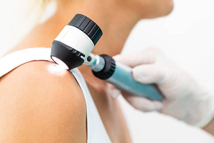
 Dermatoscope is a portable equipment that enables physician to view cuts on the skin at intensifications of around 10 times that of usual vision. It is utilized to detect and distinguish benign from malignant cuts. It is an exceptional tool for the estimate of pigmented) spots and is particularly important in identifying melanoma and other skin tumors. Utilizing dermoscopy in grouping with medical examination, palpation and the individual’s history is important to the impost of skin abrasions.
It should not be seen as an extra or second-level analytical tool to be used only in cases that are tough to analyze by medical inspection alone. Moderately, it is an essential adjunct to the medical survey of any cut that may be bothering. It can be helpful for the diagnosis of a vast range of cuts such as melanoma, squamous cell carcinoma, psoriasis, warts and other benign developments. It has also been exhibited to help in the detection of vascular series in skin lesions. It is also being utilized in the monitoring of nonpigmented cuts and is progressively recognized as a device for detecting premalignant alterations in nevi and melanocytic cancers. There are various kinds of Dermatoscopes; older designs need the usage of a linkage gel or immersion liquid such as oil, mineral oil, ultrasound gel or 70% alcohol-based commercial methods to aid illumine subsurface skin structures. Currently, a new group of dermoscopes that do not need this linkage gel have been advanced. They have an in-built polarized light which aids to decrease glare and enable for the imagining of subsurface constructions without the necessity for an immersion liquid. The Dermatoscope is an incredible device that enables providers to observe a lesion under intensification. It discloses the pigment and vascular series of the cut that can’t be seen with the naked eye. It aids to assure a diagnosis, enabling the physician to be confident in the cure of the lesion. It can also aid detect whether a cut is malignant and can forecast how the cut might change with time. This device is enormously helpful in the evaluation of any cut that break the sequence of the individual’s medical lesions - by being vast, varied, or both. While a cut can be analyzed with the naked eye, it is more precise to analyze it utilizing the dermoscopic view of the cut. It can also be utilized to aid with the resolution as to whether a cut should be detached or observed. A Dermatoscope is a portable equipment that offers light and magnifying power. It can captivate and record digital pictures, or video. The polarized light enables the provider to see from the skin surface to envisage pigment, vascular, and infectious patterns and the underlying epidermal and dermal construction of the cut. Some dermoscope can be linked to the individual’s electronic health records, enabling the doctor to upload digital pictures for observation and assessment. The dermoscope has been an important diagnostic device in the evaluation of melanoma and other benevolent nevi. It is particularly helpful in assessing nonconforming nevi and in detecting the suitability of biopsy. It can also aid doctors in gynecology, ophthalmology, urology, plastic surgery, and dentistry, and individuals doing self-examinations, to identify concerning cuts. Training medical scholars in the usage of the Dermatoscope has exhibited to enhance their awareness of atypical cuts and can reduce the number of unwanted biopsies and field referrals. While dermoscope provide advantages in the analysis of skin cancer, they are not without their limits. Some problems are difficult to identify compared to others, and the accurateness of a dermoscopic imaging can based on user skill and training, individual characteristics, and skin cut appearance.
0 Comments
Leave a Reply. |
AuthorWrite something about yourself. No need to be fancy, just an overview. Archives
July 2023
Categories
All
|
 RSS Feed
