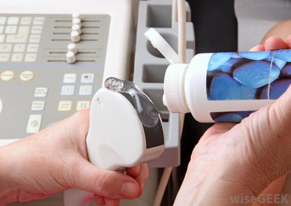
 Ultrasound Gels are a key component of ultrasound imaging, used to improve the quality of images obtained during ultrasound examinations. These gels are designed to help transmit ultrasonic waves from the transducer to the patient's skin, allowing the imaging system to capture detailed images of internal organs and tissues.
Gels are typically made from a mixture of water, glycerin, and a thickening agent, such as carbomer or xanthan gum. Some gels may also contain preservatives or other additives to enhance their performance or extend their shelf life. The main purpose of these ingredients is to help create a smooth, consistent layer of gel on the patient's skin, which allows the ultrasonic waves to travel more easily through the skin and into the underlying tissues. The glycerin in the gel helps to reduce friction between the transducer and the skin, while the thickening agent helps to keep the gel in place and prevent it from dripping or running. Ultrasound Gels are an essential part of ultrasound imaging, as they help to improve the quality of images obtained during ultrasound examinations. Without a sufficient layer of gel, the ultrasonic waves would have difficulty penetrating the skin and reaching the underlying tissues, resulting in poor-quality images that may be difficult or impossible to interpret. By providing a smooth, consistent layer of gel on the skin, gels help to ensure that the ultrasonic waves are transmitted effectively, resulting in clear, detailed images that can be used to diagnose a wide range of medical conditions. Ultrasound Gels are used in a variety of medical settings, including hospitals, clinics, and doctor's offices, and are essential for many different types of ultrasound examinations, from prenatal ultrasounds to imaging of the liver, kidneys, and other internal organs. Gels are typically applied directly to the patient's skin before the ultrasound examination. The gel is dispensed from a bottle or tube and applied to the skin in a thin, even layer, using the transducer or a gloved hand to spread it over the skin. The amount of gel required will depend on the size of the area being imaged, with larger areas requiring more gel to ensure adequate coverage. During the ultrasound examination, the transducer is placed directly on the skin, with the gel helping to create smooth, even contact between the transducer and the skin. The transducer is then moved over the skin, with the ultrasonic waves transmitted through the gel and into the underlying tissues. As the waves bounce off the internal structures of the body, they are captured by the imaging system and used to create a detailed, real-time image of the area being examined. After the examination is complete, the gel is typically wiped off the patient's skin using a damp cloth or paper towel. Some Ultrasound Gels are water-soluble and can be easily rinsed off with water, while others may require a specialized cleaning solution to remove. While gels are generally safe and well-tolerated, there are some potential risks associated with their use. Some people may experience an allergic reaction to one or more of the ingredients in the gel, such as glycerin or the thickening agent. Symptoms of an allergic reaction may include itching, hives, or difficulty breathing. In rare cases, Ultrasound Gels may cause skin irritation or redness, particularly if the gel is left on the skin for an extended period of time. Gels can become contaminated with bacteria or other microorganisms if they are not properly stored or handled. This can potentially lead to infections or other health problems.
0 Comments
Leave a Reply. |
AuthorWrite something about yourself. No need to be fancy, just an overview. Archives
July 2023
Categories
All
|
 RSS Feed
